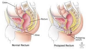
Rectal prolapse is a condition that involves the rectum turning inside out on itself and coming out through the anus. Full-thickness rectal prolapse involves the whole wall of the bowel turning inside out on itself, partial thickness involves the inner lining only.
Symptoms:
The prolapse produces an uncomfortable lump through which is red-purple. The lump may spontaneously reduce. The prolapse also causes bleeding with mucus and symptoms of incontinence.
Cause:
There are various anatomical features that may predispose: Deep anterior pelvic cul de sac, redundant sigmoid colon, diastasis of the levator ani. Rectal prolapse occurs mainly in females the peak incidence is in the 6th to 7th decade.
Investigations:
- Bedside examination including sigmoidoscopy can diagnose the condition. Examination with the person straining on the commode can diagnose it.
- Colonoscopy: usually performed in the elderly to exclude other problems.
- Examination under anaesthetic: may be needed to diagnose the problem.
- Anorectal Physiology and USS: Often used to record sphincter muscle status especially if incontinence is a major symptom.
Treatment:
In children with rectal prolapse procedures to avoid straining and holding the prolapse back in may cure the problem.
In adults with full-thickness prolapse surgery is generally required.
Over 100 different operations have been described to treat rectal prolapse.
Transabdominal Procedures:
Rectopexy alone or resection rectopexy both of which can be done laparoscopically or open.
Laparoscopic Ventral Rectopexy is a procedure now being offerred which uses a mesh placed between rectum and vagina sutured to these structures then fixed to pre-sacral tissues. This has a high success rate and is tolerated well in the elderly population. The operation avoids post-operative constipation which has been a problem in some other surgeries.
Perineal Procedures:
The Delorme procedure which is a mucosal resection and re-anastomosis, and the Altemeier or rectosigmoidectomy are the 2 commonest procedures.
Improvement in incontinence can be expected in 50% of cases.
Related Links:
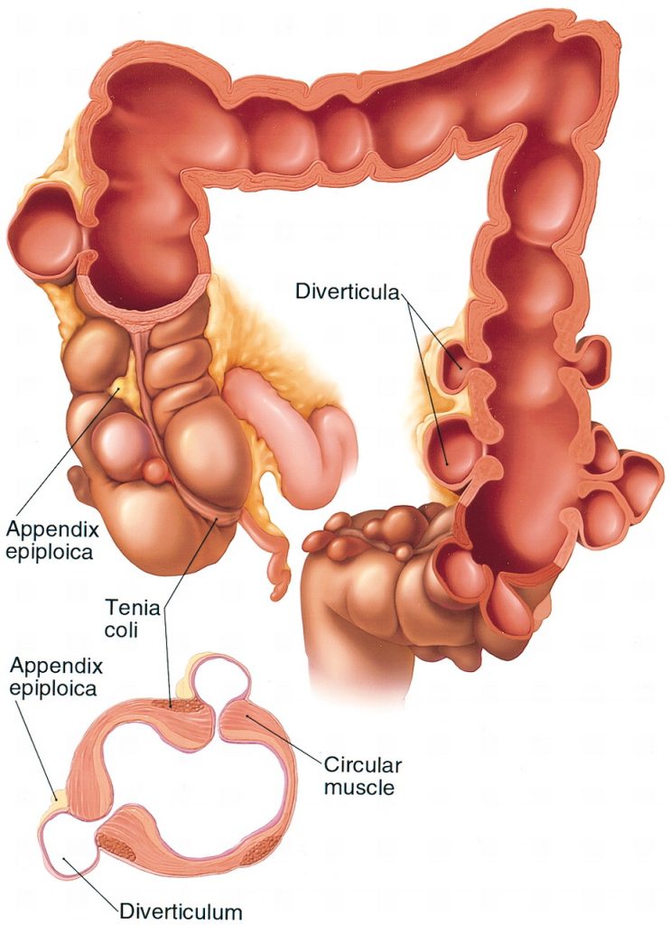
This is a common condition in our community (affecting over 50% of those over 60 years old). The colon develops weak areas in its wall which form diverticula or pockets. The sigmoid colon is the area most frequently affected and is the site of most complications.
The cause has been attributed to a chronic low fibre diet leading to increases in pressure within the colon to generate progression of stool.
Symptoms:
The condition should not cause symptoms. Left sided abdominal pain and altered bowel habits may be early symptoms. Complications cause their own specific symptoms.
Complications:
- Inflammation-Diverticulitis – causes left sided abdominal pain, and fevers
- Perforation – can cause sudden severe pain and collapse. This is a surgical emergency and hospital treatment should be obtained immediately
- Abscess – caused by a localised perforation causes ongoing pain and fevers, may cause urinary frequency and a mass can sometimes be felt.
- Fistulae – can occur especially to the bladder which may cause urinary infection or gas in the urine. In the female fistulae to the vagina can also occur causing gas and faeces to pass from the vagina.
- Large Bowel Obstruction – if scarring and muscle thickening is severe the inside can narrow and obstruct causing pain and distension.
- Bleeding – can be severe from diverticla openings and generally requires in-hospital treatment and resuscitation.
Investigations:
- Colonoscopy: often performed, diverticula openings can be seen.
- Barium enema:(X-Ray with dye and air inserted into rectum) gives a good outline of the diverticula and can show stricturing of the bowel.
- CT Scan: Can outline any mass(abscess) outside the bowel
- Nuclear Medicine Red Blood Cell Labelled scan: to localise bleeding point
- Angiogram: To localise bleeding point
- CT Angiogram: To localise bleeding point.
Treatment:
High fibre diet is the only treatment that can be offered in the setting outside acute complications
Acute infections require antibiotics and usually hospital-based observation.
Complications aside from haemorrhage generally require surgical excision of the affected segment of bowel, which can be done with open or laparoscopically assisted techniques.
Related Links:
www.fascrs.org/patients/disease-condition/diverticular-disease-expanded-version-0
www.cssanz.org/index.php/patients/diverticular-disease
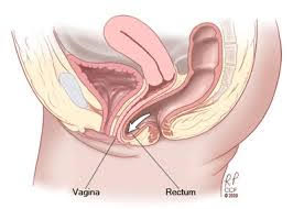
A rectocele is a weakness in the tissues of the anterior wall of the rectum and the posterior wall of the vagina and its intervening tissues. The rectal wall may bulge into the vagina particularly with straining.
Cystocele (bladder bulging into vagina), enterocele (bowel bulging into vagina), uterine prolapse can be associated conditions.
Symptoms and Signs:
- A bulge into the vagina
- Constipation and difficult defecation
- A sensation of pelvic or rectal pressure
- Incomplete evacuation
Cause:
Rectoceles often develop secondary to childbirth where the fascia between rectum and vagina are irreversibly stretched. Chronic straining such as constipation, heavy lifting, and coughing can predispose this condition. Ageing and previously having had a hysterectomy are risk factors in developing a rectocele.
Diagnosis & Investigations:
- Pelvic examination whilst straining to assess the extent of the rectocele.
- Defecating Proctogram outlines the size of the rectocele, and whether it empties during evacuation.
Treatment:
A rectocele only needs treatment if it causes a condition called obstructed defeacation whereby efforts at emptying the rectum during defeacation are impaired by stool being held up in the rectocele.
Surgical treatment in the form of rectocele repair which can be performed via the vaginal wall (transvaginal repair) or the rectal wall (transrectal) or can be performed by incising tissue between the vagina and rectum and repairing or placing mesh between the vagina and rectum.
Most series report successful results in up to 80% of cases.
A new procedure, Stapled transanal rectal resection (STARR) is currently being evaluated for use in rectocele with obstructed defeacation. The results of this procedure are good but it is too early to recommend this procedure outside a trail setting.
Laparoscopic approaches have been used in this condition but this is usually combined with treatment of other conditions such as pelvic organ prolapses and is generally not used to treat rectocele in isolation.
Links:
www.fascrs.org/patients/disease-condition/rectocele-expanded-information